By Riverwalk Dental Arts | April 2, 2026
When people think about a dental visit, they often picture the chair, the cleaning, and the exam. What they do not always see is the quiet work that helps a dentist understand what is happening beneath the surface. At Riverwalk Dental Arts in Rock Hill, that work includes digital radiography and CBCT-based 3D imaging, along with other modern tools like digital impressions and intraoral cameras. The practice lists these technologies on its Rock Hill dental technology page as part of its approach to more precise, comfortable care.
For patients, this matters because dental problems do not always announce themselves clearly. Some cavities, infections, bone changes, root issues, and jaw concerns can hide where a regular look in the mirror will never reach. The ADA says radiographs help dentists evaluate and definitively diagnose many oral diseases and conditions, but they should be used based on each patient’s needs and clinical exam. The FDA and ADA also emphasize that dental imaging should be ordered only when it is necessary for diagnosis or treatment, with benefits weighed against radiation risk.
Why 3D imaging and digital X-rays matter
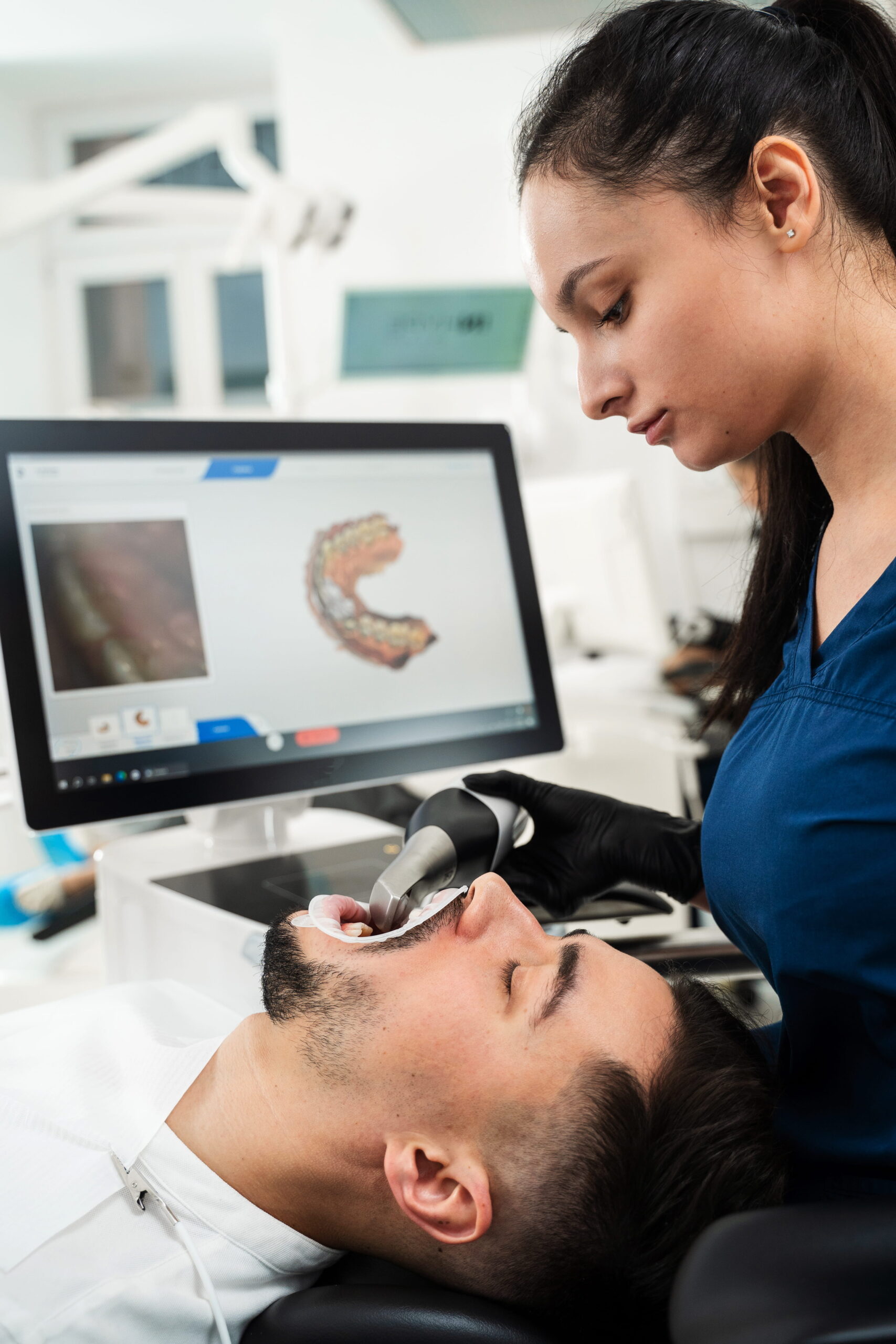
Traditional dental X-rays can show important information, but they are two-dimensional. 3D imaging, including CBCT, gives the dental team a fuller view of teeth, bone, nerve pathways, and sinus anatomy. The FDA explains that CBCT rotates around the patient and reconstructs a 3D image of the teeth and oral-maxillofacial region. It can support diagnosis, treatment planning, and evaluation for conditions such as implant planning, abnormal teeth, jaw and face concerns, root canal diagnosis, and dental trauma. Riverwalk Dental Arts describes CBCT as a way to capture precise 3D views for accurate diagnosis and personalized care.
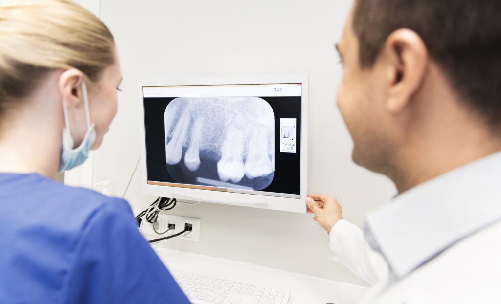
Digital radiography offers a different kind of clarity. Instead of film, it uses electronic sensors and computer systems to capture images of teeth, bone, and surrounding tissues. Riverwalk Dental Arts notes that digital images appear instantly, can be adjusted for brightness and contrast, and may reduce the need for repeat exposures. That means the dentist can move more efficiently from image to explanation, which can help patients feel more informed and less uncertain.
What patients gain from modern imaging
For many Rock Hill patients, the biggest benefit is not the technology itself. It is the confidence that comes from seeing a clearer picture before treatment begins. The ADA notes that radiographs can help dentists diagnose disease, but they should be used only when they will affect care, and the decision should be based on the patient’s oral health condition, age, disease risk, symptoms, and history. That is a thoughtful standard, not a one-size-fits-all rule.
In practical terms, modern imaging may help with:
- catching hidden decay before it becomes a bigger problem,
- evaluating infections or root issues,
- planning dental implant placement,
- checking bone levels,
- assessing jaw relationships,
- and confirming details that would be difficult to see with a basic visual exam alone.
This is especially valuable in cases where treatment needs to be exact. The more clearly the dentist can see the landscape, the better the plan can be shaped around the patient’s actual needs. That is the kind of care that reflects balance: enough information to make a sound decision, without unnecessary exposure or guesswork. The ADA’s guidance repeatedly emphasizes ALARA principles, meaning radiation exposure should be kept as low as reasonably achievable, while still supporting good diagnosis and treatment.
Is 3D imaging safe?
This is one of the most common questions patients ask, and it deserves a clear answer. The FDA says dental CBCT should be performed only when necessary to provide clinical information that cannot be provided by other imaging methods. It also notes that CBCT generally gives more radiation than conventional dental X-rays, even though it is still used for clinically important reasons and only when the expected benefit outweighs the risk. The ADA similarly advises dentists to justify every X-ray exposure and keep doses as low as reasonably achievable.
That does not mean patients should fear imaging. It means imaging should be used wisely. A careful dental team considers your history, your symptoms, and the question that needs answering before taking any image. The FDA/ADA guidance is built around that exact idea: use the right image, for the right reason, at the right time.
Why Rock Hill patients may notice the difference
Riverwalk Dental Arts positions its technology as part of a patient-centered experience. On the practice site, the team says its goal is to use technology thoughtfully to support clear diagnoses, safe treatment, and patient comfort. Its technology page also highlights CBCT, digital impressions, digital radiography, intraoral cameras, and laser dentistry, which suggests a modern workflow designed to support precision and communication.
That matters locally because patients often want care that feels efficient without feeling rushed. Clear images can shorten the distance between question and answer. They can also help patients understand what the dentist sees, which builds trust. When a dental team can point to a specific area on the screen and explain what it means, the conversation becomes easier to follow and easier to act on.
When your dentist may recommend digital X-rays or 3D imaging
Not every patient needs the same images at the same time. The ADA says there is no “one size fits all” schedule for radiographs. Instead, the dentist should consider the patient’s condition, risk factors, age, and symptoms before deciding what imaging is needed. The FDA similarly says dental CBCT should be used when other imaging cannot provide the clinical information required.
You may be more likely to need imaging if you have:
- tooth pain or swelling,
- suspected decay between teeth,
- a cracked tooth,
- a possible infection,
- gum and bone concerns,
- an implant consultation,
- or a treatment plan that needs detailed anatomical mapping.
The best imaging choice depends on the question being asked. A small concern may only need a standard digital X-ray. A more complex case may call for 3D CBCT so the dentist can see the area from multiple angles. That measured approach protects patients while supporting better clinical decisions.
What to expect during the visit
Most patients find digital imaging simple and quick. Digital X-rays use sensors instead of film, and the images appear on-screen right away. CBCT scans are also typically fast and noninvasive, with the machine rotating around the patient to create the 3D image. Riverwalk Dental Arts presents both as part of a smoother, more comfortable diagnostic experience.
Your dentist may review the images with you during the appointment, explain what they show, and connect those findings to next steps. That can make treatment feel less abstract. Instead of asking patients to trust invisible assumptions, the imaging provides something concrete to discuss.
A patient-centered way to think about dental technology
Good dental technology should not replace human judgment. It should support it. The ADA and FDA both frame radiographic guidance that way: as a resource for professional judgment, not a rigid rulebook. Riverwalk Dental Arts reflects that same balance by describing its goal as thoughtful use of technology for clearer diagnoses, safer treatment, and patient comfort.
For patients in Rock Hill, that is the real value of digital X-rays and 3D imaging. They help the dentist see more clearly, explain more clearly, and plan more carefully. In the long run, that can mean less uncertainty and more confidence in the care you receive.
FAQs
What is the difference between digital X-rays and 3D imaging?
Digital X-rays are two-dimensional images captured with electronic sensors, while CBCT creates three-dimensional images of the teeth and surrounding anatomy. CBCT can show structures from multiple angles and is often used when more detail is needed for diagnosis or treatment planning.
Are digital dental X-rays safer than traditional film X-rays?
Digital systems are designed to support efficient imaging and may reduce the need for repeat exposures. The ADA also recommends that dentists minimize radiation and order X-rays only when necessary for diagnosis or treatment.
Why would a dentist recommend CBCT?
The FDA says CBCT may help with implant planning, abnormal teeth, jaw and face evaluation, root canal diagnosis, dental trauma, and other cases where 3D information is clinically useful.
Does every patient need 3D imaging?
No. The ADA says imaging should be based on each patient’s needs, health history, risk, and symptoms. The FDA says CBCT should be used only when necessary and when other imaging methods cannot provide the needed information.