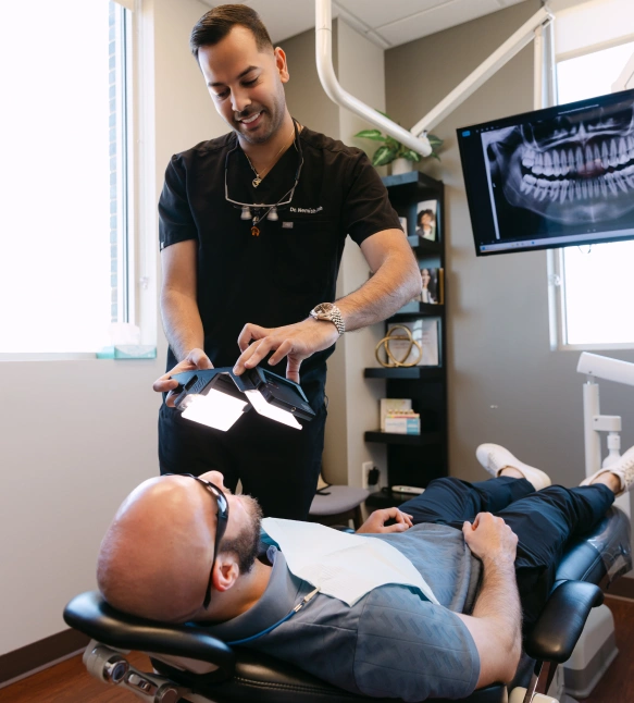
Intraoral Camera
in Rock Hill, SC

What an Intraoral Camera Does
An intraoral camera is a compact, pen-like device designed to capture clear, close-up images from inside the mouth. Its small tip and high-resolution sensor allow dental professionals to photograph teeth, gums, and other oral tissues from angles that are difficult to assess with the naked eye. The images are displayed in real time on a monitor, giving both clinician and patient a shared, magnified view of oral structures.
Unlike traditional mirrors and visual inspection alone, the intraoral camera produces full-color photos that reveal fine details such as hairline cracks, early decay, worn restorations, and subtle soft-tissue conditions. These visual records are sharp enough to be reviewed frame by frame, making it easier to pinpoint the exact location and nature of a problem. For routine exams and targeted assessments alike, the camera enhances what clinicians can see and document.
Because images are captured and stored digitally, they become part of a patient's permanent chart and can be referenced in future visits. That continuity helps clinicians track the progression of conditions over time and supports informed treatment planning. High-quality intraoral photography is now a routine component of modern dental diagnostics for its clarity and reliability.
Beyond still images, many intraoral systems offer short video capture and on-screen annotation. These features let clinicians highlight areas of interest during consultations and create a more complete visual narrative of a patient’s oral health, improving both record-keeping and clinical communication.
Educating Patients Through Real-Time Imaging
One of the most practical benefits of the intraoral camera is its role as a communication tool. When patients can see what the dentist sees, abstract explanations become concrete. Visual evidence helps patients understand the nature of a concern, the urgency of an issue, or why a particular treatment is recommended. This shared view supports clearer conversations and reduces confusion.
Seeing their own dental images on a screen empowers patients to participate more actively in decisions about their care. Clinicians can zoom, point, and annotate images to explain anatomy, demonstrate treatment needs, and outline preventive strategies. For many patients, this visual approach increases confidence in the diagnosis and fosters a collaborative relationship with the dental team.
In the context of informed consent, intraoral photography serves as an objective reference. It gives patients a factual basis for discussion and helps them remember what was shown during the appointment. This transparency supports better follow-through, whether the next steps are monitoring, hygiene-focused care, or restorative treatment.
How Intraoral Images Upgrade Clinical Records and Collaboration
Digital images from intraoral cameras integrate smoothly with modern dental record systems. Captured photos can be labeled, dated, and attached to specific chart entries so that every clinical note has a visual complement. That integration enhances clarity for the treating dentist and any specialists who may later review the case.
When collaboration is needed — for example, with an endodontist, periodontist, or dental laboratory — shared intraoral images provide a precise visual reference. A photograph can clarify margins, occlusal contacts, or soft-tissue conditions far better than a written description alone. This improves efficiency and reduces the risk of miscommunication between providers.
Images are also useful for internal quality control and treatment verification. Clinicians can document the condition of a restoration before and after care or confirm the fit of a prosthesis using photographs. Because the images are part of the electronic file, they support clinical decision-making, treatment continuity, and professional accountability.
When and Why Dentists Use an Intraoral Camera
Dentists rely on intraoral cameras during routine exams, targeted evaluations, and pre-treatment consultations. They are particularly helpful when assessing incipient decay, evaluating cracks and fractures, documenting wear patterns, or inspecting margins around crowns and fillings. The camera’s ability to reveal minute details makes it a valuable screening tool for problems that might be missed with visual inspection alone.
Intraoral imaging is also commonly used to monitor soft-tissue health. Lesions, areas of inflammation, and healing progress can be photographed and compared across visits. That consistent photographic record helps clinicians decide whether a condition warrants closer observation, biopsy, or referral to a specialist.
Because the device is noninvasive and quick to use, it fits naturally into the flow of a standard dental appointment. Captured images can guide immediate decisions during the visit, and when needed, clinicians can schedule follow-up care with a clear visual rationale to support treatment recommendations.
What Patients Can Expect During an Intraoral Camera Exam
An intraoral camera examination is typically brief and comfortable. The clinician or hygienist will gently guide the camera around the mouth while the patient remains seated. Most patients feel only minimal contact as the camera captures a series of focused images of individual teeth, gum margins, and other areas of interest.
As images appear on the monitor in real time, the clinician will point out notable findings and explain what the pictures show. This immediate feedback makes it easier to discuss preventive steps or plan restorative care. Patients are encouraged to ask questions while viewing the images, helping to demystify unfamiliar conditions.
Because the images are stored digitally, clinicians can review them later or compare them with past photographs to observe changes. This continuity supports long-term oral health planning and helps patients see the impact of their home care and clinical treatments over time.
Summary: Intraoral cameras provide a detailed, visual window into oral health that strengthens diagnosis, patient education, and clinical documentation. By combining high-resolution images with seamless digital record-keeping, this technology helps dental teams deliver clearer explanations and more confident care. If you’d like to learn how intraoral imaging is used in our practice, please contact Riverwalk Dental Arts for more information and to discuss how it may benefit your next visit.

