Digital Radiography
in Rock Hill, SC
What Digital Radiography Means for Your Dental Care
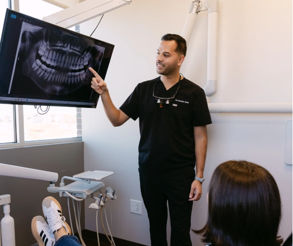
Digital radiography replaces traditional film with sensitive electronic sensors and computer systems to capture images of teeth, bone and surrounding tissues. For patients, the most noticeable difference is speed: images appear on-screen within seconds, allowing your dental team to review results with you in real time. This immediacy helps turn an appointment into a collaborative discussion rather than a waiting game.
Beyond convenience, digital imaging changes how decisions are made. Clear, high-resolution images let clinicians detect early signs of decay, bone loss, or other issues that might be missed or delayed with older methods. Because the images can be adjusted for contrast, brightness and magnification, the team can scrutinize fine details without exposing patients to repeat exposures.
We place a strong emphasis on using technology to improve the patient experience. When your visit includes radiographs, the process is designed to be efficient and minimally disruptive — you leave with a clearer understanding of your condition and the recommended next steps.
How Modern Sensors and Workflow Improve Accuracy
Instead of film, digital systems use compact sensors that capture X-ray data electronically. These sensors feed images directly into the practice’s imaging software where they are archived in the patient’s digital file. Because the transfer is instantaneous, there’s no waiting for film development and no risk of lost or damaged negatives.
Digital workflows also reduce human error. Images are automatically labeled, time-stamped and associated with the correct record, which streamlines charting and treatment planning. When images are taken chairside, clinicians can immediately verify positioning and, if necessary, retake a photo while the patient is still comfortably positioned.
Integration with other digital tools is another advantage. Images can be viewed alongside intraoral scans, clinical photographs, and CBCT exams where relevant, giving a more complete picture of oral health and enabling more precise, coordinated care.
Diagnostic Benefits That Support Better Treatment Decisions
High-quality digital images make it easier to identify problems early, which often leads to less invasive and more predictable treatments. Subtle changes in tooth structure, small cavitations, or early signs of bone loss are more readily apparent when images are sharp and adjustable. This helps the clinician prioritize issues and develop a treatment plan tailored to the patient’s needs.
Digital radiography also supports treatment documentation. Because images are stored electronically, clinicians can track changes over time and compare images side-by-side to evaluate how a condition is responding to treatment. This historical perspective is especially useful for periodontal monitoring, root canal follow-ups, and assessing restorative margins.
When complex procedures are required, these images assist in planning. Clear radiographic data helps determine the position of roots, the relationship of teeth to anatomical structures, and the extent of underlying pathology — all information that contributes to safer, more efficient care.
Patient Safety and Comfort at the Center of Imaging
One of the primary benefits of digital radiography is a substantial reduction in radiation exposure compared with older film techniques. Modern sensors are more sensitive, which means diagnostically useful images can be captured with lower doses. Reduced exposure is an important consideration for routine exams as well as for patients who require more frequent monitoring.
Comfort is improved too. Sensors are slim and designed to fit more naturally in the mouth, which decreases gag reflex and makes positioning less intrusive. The ability to see images immediately also reduces anxiety — clinicians can explain findings on the spot and answer questions while the information is fresh and visible.
Our clinical team follows established safety protocols, including proper shielding and positioning, to ensure imaging is performed responsibly. The aim is to gather the diagnostic information needed while minimizing any discomfort or risk to the patient.
Secure Management and Environmentally Responsible Care
Digital radiography eliminates chemical processing and physical film, removing hazardous waste from the imaging workflow and reducing the practice’s environmental footprint. Because images are stored electronically, there is no need for physical storage space or film archiving, which simplifies record-keeping and reduces material use.
Electronic storage also enhances data security and continuity of care. Images are saved in the patient’s chart and can be retrieved quickly for review or shared securely with specialists when coordinated care is required. When sharing is necessary, the process is handled through secure clinical channels to protect patient privacy.
These efficiencies—less waste, better organization and secure sharing—benefit both patients and the team. They keep appointments moving smoothly and make it easier to maintain accurate, comprehensive records over the long term.
What to Expect During an Imaging Appointment
When radiographs are indicated, your appointment will typically include a brief explanation of why the images are needed and what information they will provide. You may be asked to wear a lead apron for added protection while the sensor is positioned in your mouth or around your head for extraoral images. The process is quick, with most intraoral images captured in moments.
After the images are taken, your clinician will review them with you. Because digital images can be enlarged and adjusted, the team can point out areas of interest and walk you through recommended next steps. This transparent approach helps you understand the rationale behind any proposed treatment.
If follow-up images are required to monitor healing or progress, digital records make scheduling and tracking more straightforward. The practice can compare new and previous images side-by-side to assess outcomes and refine care plans as needed.
At Riverwalk Dental Arts, our goal is to use technology thoughtfully to support clear diagnoses, safe treatment, and patient comfort. If you have questions about digital radiography or how imaging fits into your dental care, please contact us for more information.