CBCT
in Rock Hill, SC

See Your Smile in 3D With CBCT Scans
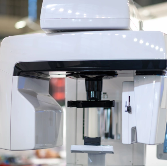
At Riverwalk Dental Arts, we combine clinical experience with modern imaging to give patients clearer answers and more predictable treatment outcomes. Three-dimensional cone-beam computed tomography (CBCT) captures detailed views of teeth, bone, nerve pathways, and sinus anatomy in ways that flat X-rays cannot, helping our team diagnose complex issues and plan treatments with greater confidence.
We prioritize tools that improve precision while keeping patient comfort and safety at the forefront of mind. CBCT imaging is fast, noninvasive, and tailored for targeted dental use—allowing our clinicians to gather the information they need without subjecting patients to lengthy procedures or unnecessary exposures.
Why 3d Imaging Matters in Modern Dentistry
Modern dentistry relies on accurate visualization. CBCT delivers a volumetric representation of the mouth and jaws, which means clinicians can view structures from any angle and examine thin slices of anatomy at high resolution. This three-dimensional perspective reduces guesswork and reveals relationships between teeth, bone, and soft tissues that are invisible on two-dimensional films.
For patients, that clarity translates into more precise diagnoses and treatment plans. Whether evaluating the extent of a lesion, assessing bone volume for restorative procedures, or investigating the source of persistent pain, CBCT equips the dental team with objective data to make informed decisions rather than relying solely on clinical impressions.
Because the scan data can be reviewed instantly and shared among specialists when necessary, treatment coordination becomes smoother. That means fewer surprises during procedures and a clearer roadmap from diagnosis to follow-up care, which is especially important for complex or multidisciplinary cases.
What CBCT Reveals That Traditional X-rays Can Miss
Traditional periapical and panoramic X-rays are valuable for many routine assessments, but they compress three dimensions into a flat image. CBCT overcomes that limitation by showing depth, cortical bone thickness, the proximity of roots to nerves, and the shape of sinus cavities. These details can change clinical decisions—such as whether a tooth can be saved, where to place an implant, or how to approach a surgical extraction.
The ability to examine thin slices through a structure also improves detection of subtle findings: small fractures, root curvatures, accessory canals, and localized bone loss can be more reliably identified. For patients experiencing unexplained symptoms, CBCT can be the diagnostic tool that uncovers underlying causes that were previously obscure.
Importantly, CBCT datasets support measurement and visualization tools used in treatment planning. Clinicians can take precise linear and volumetric measurements, annotate areas of concern, and simulate interventions. That level of detail helps set realistic expectations and informs discussions about risks and benefits.
Planning Implants and Surgery With Millimeter Accuracy
One of CBCT’s most transformative roles is in surgical planning. When placing dental implants or performing oral surgery, the difference of a few millimeters can affect function, longevity, and safety. CBCT enables exact mapping of bone height, width, and density so that implant size and position are chosen to optimize both stability and esthetics.
Surgeons also use CBCT to identify critical anatomic landmarks—such as the inferior alveolar nerve or the maxillary sinus—so surgical approaches can avoid complications. When appropriate, the imaging data can be integrated with guided-surgery protocols to fabricate surgical guides that translate the digital plan into precise clinical execution.
For patients, that means shorter chair time, fewer intraoperative surprises, and a higher likelihood of predictable healing. Clear preoperative visualization improves communication with patients about what to expect and allows for tailored post-operative care based on the exact nature of the procedure performed.
How CBCT Fits Into a Digital Dental Workflow
CBCT data integrates seamlessly with other digital technologies used in contemporary dental care. When combined with intraoral scans, CAD/CAM design, and digital planning software, the three-dimensional dataset becomes the foundation for restorations, surgical guides, and orthodontic simulations. This interoperability enhances accuracy and shortens the path from planning to final restoration.
Digital models derived from CBCT can be reviewed collaboratively by the treating dentist, laboratory technicians, and specialists, promoting a coordinated approach. For patients, this means fewer appointments, clearer visual explanations of treatment options, and a more predictable timeline for results.
Because the files are stored digitally, they also serve as a record that can be revisited for future care. That continuity supports long-term treatment planning, enabling the dental team to track changes over time and adjust strategies as needed.
In summary, cone-beam computed tomography gives dental clinicians a powerful, targeted way to visualize oral anatomy, plan procedures with greater certainty, and coordinate care across disciplines. Used thoughtfully, CBCT improves diagnostic accuracy while maintaining attention to patient safety and comfort. Contact us to learn more about how CBCT may play a role in your dental care and to discuss whether advanced 3D imaging is appropriate for your situation.
Safety, Radiation Dose, and Patient Comfort
Radiation safety is an important consideration with any imaging modality. CBCT systems designed for dental use typically deliver focused exposures and can be adjusted for the smallest possible field of view that captures the area of interest. That target-specific approach helps minimize dose while still providing diagnostic-quality images.
Providers follow the principle of using the lowest dose necessary for the clinical question at hand. Modern scanners also complete scans quickly—often within a single breath-hold—reducing motion artifact and ensuring a comfortable, stress-free experience for patients who may be anxious about imaging procedures.
Before ordering a CBCT scan, clinicians weigh the diagnostic benefit against any exposure risk and choose alternative imaging when it is sufficient. This measured approach ensures that CBCT is used judiciously as a problem-solving tool rather than a routine test for every visit.

